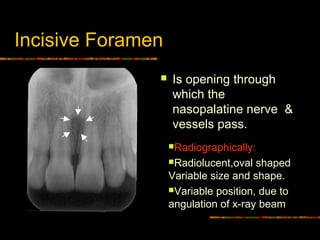
incisive foramen radiograph
Anterior extent of maxillary sinus Radiograph Frame 25. Mean canal length was 1863 235 mm and males have significantly longer incisive canal than females.

Opg Showing Incisive Foramen And Mental Foramen Download Scientific Diagram
Incisive foramen RadiographPhotograph Frame 22.
. Individual gender age race assessing technique used and degree of edentulous alveolar bone atrophy largely influence these variations. It is located in the maxilla in the incisive fossa midline in the palate posterior to the central incisors at the junction of the medial palatine and incisive sutures. It can be single or multiple.
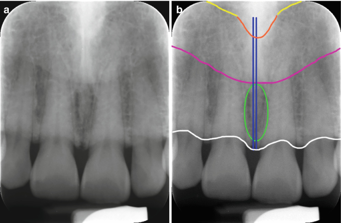
On periapical x-ray images the incisive foramen is located in the midline between the roots of the central incisors. Superior foramina of the Nasopalatine canal appear as two small rounded radiolucencies located superior to the apices of the maxillary central incisors 21. Its appearance is quite variable due to normal anatomic variation and due to the operators angulation of the x-ray beam.
Round oval lobular or heart-shaped depending on the superimposition of the anterior nasal spine 20. The incisive foramen is situated within the incisive fossa of the maxilla. The mean endpoint was approximately 1098 and 1026 mm anterior to the mental foramen for left and right side respectively without a.
However complications may arise due to an extension anterior to the mental foramen that forms the mandible incisive canal MIC. On radiographs the incisive fossa appears as a central radiolucency between the roots of the central incisors. What is the nasopalatine incisive foramen Click card to see definition.
Light green ellipsenasal openings of nasopalatine ducts. A The incisive foramen also called nasopalatine oranterior palatine foramen Fig. It gives passage blood vessels and nerves.
The mean width of the foramen labiopalatally and mesiodistally was 312 094 mm and 323 098 mm respectively. The region between mental foramens is considered as a zone of choice for implants. Inverted Y formation Radiograph comparison Frame 27.
The following characteristics of incisive were evaluated. Incisive Foramen Dr. Our goal is to evaluate identification of MIC by both panoramic radiograph PAN and cone-beam computed tomography CBCT.
Click again to see term. However complications may arise due to an extension anterior to the mental foramen that forms the mandible incisive canal MIC. Transmit nasopalatine nerves and branches of the descending palatine artery.
This radiolucency may be. In the human mouth the incisive foramen also known as. Lateral canals on each side of the midline.
On a maxillary periapical radiograph the incisive foramen appears as a small ovoid or round radiolucent area located between the roots of the maxillary central incisors 20. Incisive foramen Nasal septum Radiograph Frame 21. White ellipselateral fossa incisive fossa.
Exit through Foramina of Stenson. Maxillary sinus Radiograph Frame 24. Coronoid process is the thin triangular-shaped process of the anterosuperior aspect of the ramus.
Blackinterface between mucosa of nasal floor and inferior nasal meatus and air in inferior nasal meatus. The region between mental foramens is considered as a zone of choice for implants. 2A 3A is seen as an oval radiolucency between the roots of the maxillary central inci- sors.
Tap card to see definition. Median palatal suture D. It suggests that the clinicians should carefully identify these.
It is actually in the anterior part of the palate but superimposition makes it appear to be located between the roots of the central incisors. Assessment of the mandibular incisive canal by panoramic radiograph and cone-beam computed tomography Objectives. Yellowfloor of nasal cavity.
Incisive foramen Median palatine suture Pterygoid plates Pterodactyl gr. 7 Landmarks in the Maxilla Anterior nasal spine Zygomatic process Pterygoid plates Coronoid process of the mandible Nasolabial fold Coronoid Process From the Greek word for Crows Beak. Maxillary sinus Illustration Frame 23.
The incisive foramen also known as nasopalatine foramen or anterior palatine foramen is the oral opening of the nasopalatine canal. 1 Width of the nasopalatine canal labiopalatally and mesiodistally Figures 1 a and 1 b 2 Length of the canal Figure 1 c 3 Width of the bone anterior to the canal Figure 1 d 4 Shape of the canal Figures 2 a 2 d. Inverted Y formation Radiograph Frame 26.
On a _____ periapical radiograph the incisive foramen appears as a small ovoid or round _____ area located between the roots of the central incisors. The mean width of bone anterior to the incisive canal was 632 143 mm. Results The incisive canal was found in 87 of the scans.
Light blueanterior recess of right maxillary sinus. Anterior palatine foramen or nasopalatine foramen is the opening of the incisive canals on the hard palate immediately behind the incisor teeth. The mandibular incisive canal mental foramen and associated neurovascular bundles exist in different locations and possess many variations.
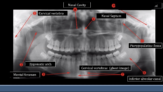
Nizar Mujallid En Twitter Interpretation Of Opg Anatomical Landmarks Https T Co Agptrcwlvs Twitter

Pin By Windy Rothmund On Dental Hygiene Butterfly Shape Sphenoid Bone Palatine

Normal Radiographic Anatomical Landmarks

Anatomical Landmarks Of Panoramic Radiographs With Ppt Lecture Note For Download Lecture Notes In Dental Assistant Study Dental Hygiene School Dentistry

Panoramic Radiograph Showing Mandibular Incisive Canal Arrow Download Scientific Diagram

Based On Ct Sagittal Sections Of The Incisive Canals The Diameters Of Download Scientific Diagram
Normal Anatomical Landmarks In Dental X Rays And Cbct Springerlink
![]()
Mandibular Incisive Canal Arrow Running From Mental Foramen Circle Download Scientific Diagram
Intra Oral Radiographic Anatomical Landmarks

Normal Anatomical Landmarks In Dental X Rays And Cbct Springerlink

Measurement Of Nasopalatine Canal Length A Incisive Foramen Diameter Download Scientific Diagram

Pdf The Evaluation Of Visibility Of Mandibular Anatomic Landmarks Using Panoramic Radiography Semantic Scholar

Mouth Incisive Canal Cyst Professional Radiology Outcomes

Audience Dental Students Intended Purpose To Be Used As Part Of A Dental Course Teaching Students About This Procedure Dental Student Dentistry Dental

An Example Of A Large Incisive Canal Mesial To The Mental Foramen The Download Scientific Diagram

The Nasopalatine Canal On Cbct Images A An Axial Section At The Level Download Scientific Diagram

Pin On Teeth

Interior Skull Skull Anatomy Axial Skeleton Sphenoid Bone

Visibility Of Mandibular Anatomical Landmarks In Panoramic Radiography A Retrospective Study Semantic Scholar
0 Response to "incisive foramen radiograph"
Post a Comment